Abstract:  Cerebral ischemia is a major health risk that requires preventive approaches in addition to drug therapy. Physical exercise enhances neurogenesis and synaptogenesis, and has been widely used for functional rehabilitation after stroke. In this study, we determined whether exercise training before disease onset can alleviate the severity of cerebral ischemia. We also examined the role of exercise-induced circulating factors in these effects. Adult mice were subjected to 14 days of treadmill exercise training before surgery for middle cerebral artery occlusion. We found that this exercise pre-conditioning strategy effectively attenuated brain infarct area, inhibited gliogenesis, protected synaptic proteins, and improved novel object and spatial memory function. Further analysis showed that circulating adiponectin plays a critical role in these preventive effects of exercise. Agonist activation of adiponectin receptors by AdipoRon mimicked the effects of exercise, while inhibiting receptor activation abolished the exercise effects. In summary, our results suggest a crucial role of circulating adiponectin in the effects of exercise pre-conditioning in protecting against cerebral ischemia and supporting the health benefits of exercise.

Zheng M, Zhang B, Yau SSY, So KF, Zhang L, Ou H. Exercise preconditioning alleviates ischemia-induced memory deficits by increasing circulating adiponectin. Neural Regen Res. 2025 May 1;20(5):1445-1454. doi: 10.4103/NRR.NRR-D-23-01101. Epub 2024 Mar 1. PMID: 39075911..
https://pubmed.ncbi.nlm.nih.gov/39075911/

Posted in Uncategorized | Comments Off on Exercise preconditioning alleviates ischemia-induced memory deficits by increasing circulating adiponectin

Abstract

Background

Bipolar Disorder (BD), a severe neuropsychiatric condition, often appears during adolescence. Traditional diagnostic methods, which primarily relying on clinical interviews and single-modal MRI techniques, may have limitations in accuracy. This study aimed to improve adolescent BD diagnosis by integrating behavioral assessments with multimodal MRI. We hypothesized that this combination would enhance diagnostic accuracy for at-risk adolescents.

Methods

A retrospective cohort of 309 subjects, including BD patients, offspring of BD patients (with and without subthreshold symptoms), non-BD offspring with subthreshold symptoms, and healthy controls, was analysed. Behavioral attributes were integrated with MRI features from T1, rsfMRI, and DTI. Three diagnostic models were developed using GLMNET multinomial regression: a clinical diagnosis model based on behavioral attributes, an MRI-based model, and a comprehensive model integrating both datasets.

Results

The comprehensive model achieved a prediction accuracy of 0.83 (CI: [0.72, 0.92]), significantly higher than the clinical (0.75) and MRI-based (0.65) models. Validation with an external cohort showed high accuracy (0.89, AUC=0.95). Structural equation modelling revealed that Clinical Diagnosis (β=0.487, p<0.0001), Parental BD History (β=-0.380, p<0.0001), and Global Function (β=0.578, p<0.0001) significantly impacted Brain Health, while Psychiatric Symptoms showed only a marginal influence (β=-0.112, p=0.056).

Conclusion

This study highlights the value of integrating multimodal MRI with behavioral assessments for early diagnosis in at-risk adolescents. Combining neuroimaging enables more accurate patient subgroup distinctions, facilitating timely interventions and improving health outcomes. Our findings suggest a paradigm shift in BD diagnostics, advocating for incorporating advanced imaging techniques.

Wu J, Lin K, Lu W, Zou W, Li X, Tan Y, Yang J, Zheng D, Liu X, Lam BY, Xu G, Wang K, McIntyre RS, Wang F, So KF, Wang J. Enhancing Early Diagnosis of Bipolar Disorder in Adolescents through Multimodal Neuroimaging. Biol Psychiatry. 2024 Jul 26:S0006-3223(24)01485-9. doi: 10.1016/j.biopsych.2024.07.018. Epub ahead of print. PMID: 39069165.

https://www.sciencedirect.com/science/article/pii/S0006322324014859


Posted in Uncategorized | Comments Off on Enhancing Early Diagnosis of Bipolar Disorder in Adolescents through Multimodal Neuroimaging

Abstract: Severe Acute Respiratory Syndrome Coronavirus 2 (SARS-CoV-2) infection inhibits mitochondrial oxidative phosphorylation (OXPHOS) and elevates mitochondrial reactive oxygen species (ROS, mROS) which activates hypoxia-inducible factor-1alpha (HIF-1α), shifting metabolism toward glycolysis to drive viral biogenesis but also causing the release of mitochondrial DNA (mtDNA) and activation of innate immunity. To determine whether mitochondrially targeted antioxidants could mitigate these viral effects, we challenged mice expressing human angiotensin-converting enzyme 2 (ACE2) with SARS-CoV-2 and intervened using transgenic and pharmacological mitochondrially targeted catalytic antioxidants. Transgenic expression of mitochondrially targeted catalase (mCAT) or systemic treatment with EUK8 decreased weight loss, clinical severity, and circulating levels of mtDNA; as well as reduced lung levels of HIF-1α, viral proteins, and inflammatory cytokines. RNA-sequencing of infected lungs revealed that mCAT and Eukarion 8 (EUK8) up-regulated OXPHOS gene expression and down-regulated HIF-1α and its target genes as well as innate immune gene expression. These data demonstrate that SARS-CoV-2 pathology can be mitigated by catalytically reducing mROS, potentially providing a unique host-directed pharmacological therapy for COVID-19 which is not subject to viral mutational resistance.


Guarnieri JW, Lie T, Albrecht YES, Hewin P, Jurado KA, Widjaja GA, Zhu Y, McManus MJ, Kilbaugh TJ, Keith K, Potluri P, Taylor D, Angelin A, Murdock DG, Wallace DC. Mitochondrial antioxidants abate SARS-COV-2 pathology in mice. Proc Natl Acad Sci U S A. 2024 Jul 23;121(30):e2321972121. doi: 10.1073/pnas.2321972121. Epub 2024 Jul 15. PMID: 39008677.


https://pubmed.ncbi.nlm.nih.gov/39008677/

Posted in Uncategorized | Comments Off on Mitochondrial antioxidants abate SARS-COV-2 pathology in mice

Summary: “Osteoarthritis (OA) is a degenerative joint disease and the most common form of arthritis globally, which is a leading cause of disability among the elderly population. The typical symptoms of OA include pain and stiffness in the affected joints. The current treatment strategies for OA include regular exercise, acupuncture, and massage. Patients with severe OA have to undergo surgery, such as joint replacement. Although the etiology of OA remains not fully understood, it is well recognized that the pathogenesis and progression of OA are closely associated with abnormal immune responses in the cartilage microenvironment (1). Apart from regulatory proteins, emerging evidence has underscored the crucial roles of immunoregulatory metabolites in various immune disorders (2). Compared to traditional medications, natural metabolites, many of which are derived from our daily diet, usually have advantages in terms of higher availability, proven safety, and lower costs. Therefore, the diet-based intervention of immunological diseases has been attracting increasingly more attention in recent years. However, the metabolic alterations in the cartilage microenvironment of OA patients and their potential effects on OA pathogenesis remain largely unexplored to date. Herein, we report a unique, anti-OA role of vitamin B1 (VB1), and the underlying mechanism was further explored.”

Shen S, Liang Y, Zhao Y, Hu Z, Huang Y, Wu Y, Liu Y, Fan S, Wang Q, Xiao P. Dietary supplementation of vitamin B1 prevents the pathogenesis of osteoarthritis. Proc Natl Acad Sci U S A. 2024 Jul 23;121(30):e2408160121. doi: 10.1073/pnas.2408160121. Epub 2024 Jul 18. PMID: 39024114.

https://www.pnas.org/doi/10.1073/pnas.2408160121

Posted in Uncategorized | Comments Off on Dietary supplementation of vitamin B1 prevents the pathogenesis of osteoarthritis

Highlights

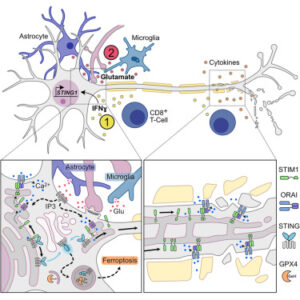
  • STING is expressed in human and mouse neurons solely under inflammatory conditions
  • Excitotoxicity activates a non-canonical STIM1-STING signaling pathway in neurons
  • Neuronal STING activation leads to autophagic GPX4 degradation and ferroptosis
  • Targeting neuronal STING protects from inflammation-induced neurodegeneration

Summary

Inflammation-induced neurodegeneration is a defining feature of multiple sclerosis (MS), yet the underlying mechanisms remain unclear. By dissecting the neuronal inflammatory stress response, we discovered that neurons in MS and its mouse model induce the stimulator of interferon genes (STING). However, activation of neuronal STING requires its detachment from the stromal interaction molecule 1 (STIM1), a process triggered by glutamate excitotoxicity. This detachment initiates non-canonical STING signaling, which leads to autophagic degradation of glutathione peroxidase 4 (GPX4), essential for neuronal redox homeostasis and thereby inducing ferroptosis. Both genetic and pharmacological interventions that target STING in neurons protect against inflammation-induced neurodegeneration. Our findings position STING as a central regulator of the detrimental neuronal inflammatory stress response, integrating inflammation with glutamate signaling to cause neuronal cell death, and present it as a tractable target for treating neurodegeneration in MS.

.

Woo MS, Mayer C, Binkle-Ladisch L, Sonner JK, Rosenkranz SC, Shaposhnykov A, Rothammer N, Tsvilovskyy V, Lorenz SM, Raich L, Bal LC, Vieira V, Wagner I, Bauer S, Glatzel M, Conrad M, Merkler D, Freichel M, Friese MA. STING orchestrates the neuronal inflammatory stress response in multiple sclerosis. Cell. 2024 Jun 7:S0092-8674(24)00576-2. doi: 10.1016/j.cell.2024.05.031. Epub ahead of print. PMID: 38878778.

https://www.cell.com/cell/fulltext/S0092-8674(24)00576-2

Posted in Uncategorized | Comments Off on STING orchestrates the neuronal inflammatory stress response in multiple sclerosis

Abstract:    Machine learning can be used to define subtypes of psychiatric conditions based on shared biological foundations of mental disorders. Here we analyzed cross-sectional brain images from 4,222 individuals with schizophrenia and 7038 healthy subjects pooled across 41 international cohorts from the ENIGMA, non-ENIGMA cohorts and public datasets. Using the Subtype and Stage Inference (SuStaIn) algorithm, we identify two distinct neurostructural subgroups by mapping the spatial and temporal ‘trajectory’ of gray matter change in schizophrenia. Subgroup 1 was characterized by an early cortical-predominant loss with enlarged striatum, whereas subgroup 2 displayed an early subcortical-predominant loss in the hippocampus, striatum and other subcortical regions. We confirmed the reproducibility of the two neurostructural subtypes across various sample sites, including Europe, North America and East Asia. This imaging-based taxonomy holds the potential to identify individuals with shared neurobiological attributes, thereby suggesting the viability of redefining existing disorder constructs based on biological factors.

Jiang Y, Luo C, Wang J, Palaniyappan L, Chang X, Xiang S, Zhang J, Duan M, Huang H, Gaser C, Nemoto K, Miura K, Hashimoto R, Westlye LT, Richard G, Fernandez-Cabello S, Parker N, Andreassen OA, Kircher T, Nenadić I, Stein F, Thomas-Odenthal F, Teutenberg L, Usemann P, Dannlowski U, Hahn T, Grotegerd D, Meinert S, Lencer R, Tang Y, Zhang T, Li C, Yue W, Zhang Y, Yu X, Zhou E, Lin CP, Tsai SJ, Rodrigue AL, Glahn D, Pearlson G, Blangero J, Karuk A, Pomarol-Clotet E, Salvador R, Fuentes-Claramonte P, Garcia-León MÁ, Spalletta G, Piras F, Vecchio D, Banaj N, Cheng J, Liu Z, Yang J, Gonul AS, Uslu O, Burhanoglu BB, Uyar Demir A, Rootes-Murdy K, Calhoun VD, Sim K, Green M, Quidé Y, Chung YC, Kim WS, Sponheim SR, Demro C, Ramsay IS, Iasevoli F, de Bartolomeis A, Barone A, Ciccarelli M, Brunetti A, Cocozza S, Pontillo G, Tranfa M, Park MTM, Kirschner M, Georgiadis F, Kaiser S, Van Rheenen TE, Rossell SL, Hughes M, Woods W, Carruthers SP, Sumner P, Ringin E, Spaniel F, Skoch A, Tomecek D, Homan P, Homan S, Omlor W, Cecere G, Nguyen DD, Preda A, Thomopoulos SI, Jahanshad N, Cui LB, Yao D, Thompson PM, Turner JA, van Erp TGM, Cheng W; ENIGMA Schizophrenia Consortium; Feng J; ZIB Consortium. Neurostructural subgroup in 4291 individuals with schizophrenia identified using the subtype and stage inference algorithm. Nat Commun. 2024 Jul 17;15(1):5996. doi: 10.1038/s41467-024-50267-3. PMID: 39013848; PMCID: PMC11252381.

https://pubmed.ncbi.nlm.nih.gov/39013848/

Posted in Uncategorized | Comments Off on Neurostructural subgroup identified in 4291 individuals with schizophrenia
OBJECTIVES:

Autism spectrum disorder (ASD) is estimated to be ∼10 times higher in children with versus without an autistic sibling in population-based studies. Prospective studies of infant siblings have revealed even higher familial recurrence rates. In the current prospective longitudinal study, we provide updated estimates of familial ASD recurrence using a multinational database of infants with older autistic siblings.

METHODS:

Data were collated across 18 sites of the Baby Siblings Research Consortium, an international network studying the earliest manifestations of ASD. A total of 1605 infants with an older autistic sibling were followed from early in life to 3 years, when they were classified as ASD or non-ASD. Hierarchical generalized linear modeling, with site as a random effect, was used to examine predictors of recurrence in families and calculate likelihood ratios.

RESULTS:

A total of 20.2% of siblings developed ASD, which is not significantly higher than the previously reported rate of 18.7%. Male infant sex and >1 older affected sibling were significant predictors of familial recurrence. Proband sex also influenced recurrence rates, with siblings of female probands significantly more likely to develop ASD than siblings of male probands. Race and maternal education were also associated with recurrence in families.

CONCLUSIONS:

The familial recurrence rate of ASD, as measured in infant sibling studies, has not changed appreciably since previous estimates were made in 2011. Younger siblings of autistic children, particularly those who are male, have an affected female sibling, multiple affected siblings, or are impacted by social inequities, should be closely monitored and promptly referred for diagnostic evaluation.

Ozonoff, S. et al.: Familial Recurrence of Autism: Updates From the Baby Siblings Research Consortium. Pediatrics (July 16, 2024) e2023065297. https://doi.org/10.1542/peds.2023-065297

https://publications.aap.org/pediatrics/article/doi/10.1542/peds.2023-065297/197777/Familial-Recurrence-of-Autism-Updates-From-the

Posted in Uncategorized | Comments Off on Familial Recurrence of Autism: Updates From the Baby Siblings Research Consortium

Editor’s summary

Although gene editing is beginning to achieve clinical acceptance as an intervention for some genetic diseases, difficulties remain. Challenges include efficient editing of cells directly in an organism rather than ex vivo and long-term persistence of the edits, which requires targeting the relevant stem cells. Sun et al. designed lipid nanoparticles for delivering gene-editing reagents and optimized them to deliver these reagents to all lung cell types, including stem cells (see the Perspective by Bulcaen and Carlon). The authors then tested their approach in patient-derived cells and in a mouse model of cystic fibrosis, demonstrating that they could achieve therapeutically relevant editing lasting for up to a year. —Yevgeniya Nusinovich

Abstract

In vivo genome correction holds promise for generating durable disease cures; yet, effective stem cell editing remains challenging. In this work, we demonstrate that optimized lung-targeting lipid nanoparticles (LNPs) enable high levels of genome editing in stem cells, yielding durable responses. Intravenously administered gene-editing LNPs in activatable tdTomato mice achieved >70% lung stem cell editing, sustaining tdTomato expression in >80% of lung epithelial cells for 660 days. Addressing cystic fibrosis (CF), NG-ABE8e messenger RNA (mRNA)–sgR553X LNPs mediated >95% cystic fibrosis transmembrane conductance regulator (CFTR) DNA correction, restored CFTR function in primary patient-derived bronchial epithelial cells equivalent to Trikafta for F508del, corrected intestinal organoids and corrected R553X nonsense mutations in 50% of lung stem cells in CF mice. These findings introduce LNP-enabled tissue stem cell editing for disease-modifying genome correction.

Sun, Y. et al: In Vivo editing of lung STEM cells for durable gene correction in mice. Science https://doi.org/10.1126/science. adk9428 (2024).

https://www.science.org/doi/10.1126/science.adk9428

Posted in Uncategorized | Comments Off on In vivo editing of stem cells for durable gene correction in mice

Abstract:

Chronic low-grade inflammation and neuronal deregulation are two components of a smoldering disease activity that drives the progression of disability in people with multiple sclerosis (MS). Although several therapies exist to dampen the acute inflammation that drives MS relapses, therapeutic options to halt chronic disability progression are a major unmet clinical need. The development of such therapies is hindered by our limited understanding of the neuron-intrinsic determinants of resilience or vulnerability to inflammation. In this Review, we provide a neuron-centric overview of recent advances in deciphering neuronal response patterns that drive the pathology of MS. We describe the inflammatory CNS environment that initiates neurotoxicity by imposing ion imbalance, excitotoxicity and oxidative stress, and by direct neuro-immune interactions, which collectively lead to mitochondrial dysfunction and epigenetic dysregulation. The neuronal demise is further amplified by breakdown of neuronal transport, accumulation of cytosolic proteins and activation of cell death pathways. Continuous neuronal damage perpetuates CNS inflammation by activating surrounding glia cells and by directly exerting toxicity on neighbouring neurons. Further, we explore strategies to overcome neuronal deregulation in MS and compile a selection of neuronal actuators shown to impact neurodegeneration in preclinical studies. We conclude by discussing the therapeutic potential of targeting such neuronal actuators in MS, including some that have already been tested in interventional clinical trials.

Woo, M.S., Engler, J.B. & Friese, M.A. The neuropathobiology of multiple sclerosis. Nat. Rev. Neurosci. 25493–513 (2024). https://doi.org/10.1038/s41583-024-00823-z

https://www.nature.com/articles/s41583-024-00823-z

Posted in Uncategorized | Comments Off on The neuropathobiology of multiple sclerosis

Abstract: “MiR-142-3p has recently emerged as key factor in tailoring personalized treatments for multiple sclerosis (MS), a chronic autoimmune demyelinating disease of the central nervous system (CNS) with heterogeneous pathophysiology and an unpredictable course. With its involvement in a detrimental regulatory axis with interleukin-1beta (IL1β), miR-142-3p orchestrates excitotoxic synaptic alterations that significantly impact both MS progression and therapeutic outcomes. In this study, we investigated for the first time the influence of individual genetic variability on the miR-142-3p excitotoxic effect in MS. We specifically focused on the single-nucleotide polymorphism Val66Met (rs6265) of the brain-derived neurotrophic factor (BDNF) gene, known for its crucial role in CNS functioning. We assessed the levels of miR-142-3p and IL1β in cerebrospinal fluid (CSF) obtained from a cohort of 114 patients with MS upon diagnosis. By stratifying patients according to their genetic background, statistical correlations with clinical parameters were performed. Notably, in Met-carrier patients, we observed a decoupling of miR-142-3p levels from IL1β levels in the CSF, as well as from of disease severity (Expanded Disability Status Score, EDSS; Multiple Sclerosis Severity Score, MSSS; Age-Related Multiple Sclerosis Severity Score, ARMSS) and progression (Progression Index, PI). Our discovery of the interference between BDNF Val66Met polymorphism and the synaptotoxic IL1β-miR-142-3p axis, therefore hampering miR-142-3p action on MS course, provides valuable insights for further development of personalized medicine in the field.”

Dolcetti E, Musella A, Balletta S, Gilio L, Bruno A, Stampanoni Bassi M, Lauritano G, Buttari F, Fresegna D, Tartacca A, Mariani F, Palmerio F, Rovella V, Ferese R, Gambardella S, Giardina E, Finardi A, Furlan R, Mandolesi G, Centonze D, De Vito F. Interaction between miR-142-3p and BDNF Val/Met Polymorphism Regulates Multiple Sclerosis Severity. Int J Mol Sci. 2024 May 11;25(10):5253. doi: 10.3390/ijms25105253. PMID: 38791290; PMCID: PMC11121620.

https://pubmed.ncbi.nlm.nih.gov/38791290/

Posted in Uncategorized | Comments Off on Interaction between miR-142-3p and BDNF Val/Met Polymorphism Regulates Multiple Sclerosis Severity