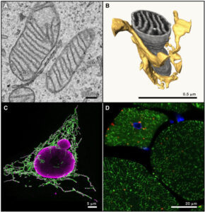
Sigma non-opioid intracellular receptor 1 (Sigma-1R) is an intracellular chaperone protein residing on the endoplasmic reticulum at the mitochondrial-associated membrane (MAM) region. Sigma-1R is abundant in the brain and is involved in several physiological processes as well as in various disease states. The role of Sigma-1R at the blood-brain barrier (BBB) is incompletely characterized. In this study, the effect of Sigma-1R activation was investigated in vitro on rat brain microvascular endothelial cells (RBMVEC), an important component of the blood-brain barrier (BBB), and in vivo on BBB permeability in rats. The Sigma-1R agonist PRE-084 produced a dose-dependent increase in mitochondrial calcium, and mitochondrial and cytosolic reactive oxygen species (ROS) in RBMVEC. PRE-084 decreased the electrical resistance of the RBMVEC monolayer, measured with the electric cell-substrate impedance sensing (ECIS) method, indicating barrier disruption. These effects were reduced by pretreatment with Sigma-1R antagonists, BD 1047 and NE 100. In vivo assessment of BBB permeability in rats indicates that PRE-084 produced a dose-dependent increase in brain extravasation of Evans Blue and sodium fluorescein brain; the effect was reduced by the Sigma-1R antagonists. Immunocytochemistry studies indicate that PRE-084 produced a disruption of tight and adherens junctions and actin cytoskeleton. The brain microcirculation was directly visualized in vivo in the prefrontal cortex of awake rats with a miniature integrated fluorescence microscope (aka, miniscope; Doric Lenses Inc.). Miniscope studies indicate that PRE-084 increased sodium fluorescein extravasation in vivo. Taken together, these results indicate that Sigma-1R activation promoted oxidative stress and increased BBB permeability.

Brailoiu E, Barr JL, Wittorf HN, Inan S, Unterwald EM, Brailoiu GC. Modulation of the Blood-Brain Barrier by Sigma-1R Activation. Int J Mol Sci. 2024 May 9;25(10):5147. doi: 10.3390/ijms25105147. PMID: 38791182; PMCID: PMC11121402.

https://pubmed.ncbi.nlm.nih.gov/38791182/
 

Posted in Uncategorized | Comments Off on Modulation of the Blood-Brain Barrier by Sigma-1R Activation

Abstract

Background: Previous studies have shown that brain volume is negatively associated with cigarette smoking, but there is an ongoing debate about whether smoking causes lowered brain volume or a lower brain volume is a risk factor for smoking. We address this debate through multiple methods that evaluate directionality: Bradford Hill’s criteria, which are commonly used to understand a causal relationship in epidemiological studies, and mediation analysis.  Methods: In 32,094 participants of European descent from the UK Biobank dataset, we examined the relationship between a history of daily smoking and brain volumes, as well as an association of genetic risk score to ever smoking with brain volume.  Results: A history of daily smoking was strongly associated with decreased brain volume, and a history of heavier smoking was associated with a greater decrease in brain volume. The strongest association was between total gray matter volume and a history of daily smoking (effect size = −2964 mm3, p = 2.04 × 10−16), and there was a dose-response relationship with more pack years smoked associated with a greater decrease in brain volume. A polygenic risk score for smoking initiation was strongly associated with a history of daily smoking (effect size = 0.05, p = 4.20 × 10−84), but only modestly associated with total gray matter volume (effect size = −424 mm3, p = .01). Mediation analysis indicated that a history of daily smoking mediated the relationship between the smoking initiation polygenic risk score and total gray matter volume. Conclusions: A history of daily smoking is strongly associated with a decreased total brain volume.

 .

Chang Y, Thornton V, Chaloemtoem A, Anokhin AP, Bijsterbosch J, Bogdan R, Hancock DB, Johnson EO, Bierut LJ. Investigating the Relationship Between Smoking Behavior and Global Brain Volume. Biol Psychiatry Glob Open Sci. 2023 Oct 6;4(1):74-82. doi: 10.1016/j.bpsgos.2023.09.006. PMID: 38130847; PMCID: PMC10733671.

https://www.bpsgos.org/article/S2667-1743(23)00136-2/fulltext

Posted in Uncategorized | Comments Off on Relationship Between Smoking Behavior and Global Brain Volume

Cow’s Milk Containing Avian Influenza A(H5N1) Virus — Heat Inactivation and Infectivity in Mice

Published May 24, 2024
DOI: 10.1056/NEJMc2405495
In late March 2024, highly pathogenic avian influenza virus (HPAI) of the H5N1 subtype was for the first time detected in nasal swabs and milk of dairy cows, increasing concern that HPAI A(H5N1) viruses may enter the human food chain. The Texas A&M Veterinary Medical Diagnostic Laboratory obtained cow’s milk samples from an affected herd in New Mexico, from which eight HPAI A(H5N1) viruses were isolated (Table S1; for details, see the Supplementary Appendix, available with the full text of this letter at NEJM.org).
We compared the genetic origin of these HPAI A(H5N1) milk virus isolates with the sequences publicly available at the time of our analysis (Fig. S1 in the Supplementary Appendix). The cow viruses form a single clade encompassing many smaller clades of viruses isolated from cats, raccoons, chickens, and wild birds. The phylogeny is consistent with a single introduction into cows. The viruses isolated in our study (labeled in Fig. S1) fall within the clade of publicly available cow virus sequences, including that from a human isolate, A/Texas/37/2024 (Fig. S1). Further assessment of the cow virus sequences and all avian influenza A virus sequences collected in the Americas since the start of 2020 identified a reassortment event for NP and PB2 segments that occurred immediately before the introduction of HPAI A(H5N1) viruses into cows (Fig. S2), consistent with findings reported by Anderson and colleagues.1
Studies involving foot-and-mouth disease virus revealed that heat inactivation of virus-positive milk samples required higher temperature or longer incubation times (or both) than heat inactivation of virus spiked into milk,2,3 presumably because fat globules and casein micelles may partly protect viruses in virus-positive milk samples. Accordingly, we tested heat inactivation of four HPAI A(H5N1) virus–positive milk samples (NM#93, NM#115, KS#3, and KS#6). Undiluted milk samples were incubated in a polymerase-chain-reaction (PCR) thermocycler at 63°C for 5, 10, 20, or 30 minutes or at 72°C for 5, 10, 15, 20, or 30 seconds (Table 1; see also the Supplementary Appendix). Control samples were left untreated. Heat treatment at 63°C reduced the virus titers below the detection limit of the TCID50 (50% tissue-culture infectious dose) assay (1.5 log10/ml). Heat treatment at 72°C was performed, with the default settings of the PCR thermocycler (i.e., preheated lid at 105°C) or with a metal lid (heated to 72°C) covering the PCR block (see the Supplementary Appendix for details). After heat treatment, samples were inoculated into embryonated chicken eggs or Madin–Darby canine kidney (MDCK) cells for virus detection. Under these conditions, heat treatment for 15 or 20 seconds reduced virus titers by more than 4.5 log units but did not completely inactivate the virus (Table 1). We emphasize that the conditions used in our laboratory study are not identical to the large-scale industrial treatment of raw milk.
The stability of HPAI A(H5N1) virus in cow’s milk stored at 4°C is another important question. For milk sample NM#93, we detected a decline of only two log units over 5 weeks. HPAI A(H5N1) virus may therefore remain infectious for several weeks in raw milk kept at 4°C.
To further assess the risk that HPAI A(H5N1)–positive milk poses to animals and humans, we orally inoculated BALB/cJ mice with 50 μl (3×10⁶ pfu) of sample NM#93. The animals showed signs of illness starting on day 1, including ruffled fur and lethargy. All the animals survived until day 4, when they were euthanized to determine virus titers in multiple organs (Figure S3 in the Supplementary Appendix). We detected high virus titers in the respiratory organs (which suggests that infection may have occurred through the pharynx) and moderate virus titers in several other organs, findings consistent with the systemic infections typically caused by HPAI H5 viruses in mammals. Detection of virus in the mammary glands of two mice was consistent with the high virus load in the milk of lactating cows, even though these mice were not lactating. Collectively, our data indicate that HPAI A(H5N1) virus in untreated milk can infect susceptible animals that consume it. In summary, HPAI H5–positive milk poses a risk when consumed untreated, but heat inactivation under the laboratory conditions used here reduces HPAI H5 virus titers by more than 4.5 log units. However, bench-top experiments do not recapitulate commercial pasteurization processes.
Lizheng Guan, Ph.D.
Amie J. Eisfeld, Ph.D.
David Pattinson, Ph.D.
Chunyang Gu, Ph.D.
Asim Biswas, Ph.D.
Tadashi Maemura, Ph.D.
Sanja Trifkovic, Ph.D.
Lavanya Babujee, Ph.D.
Robert Presler, Jr., M.S.
Randall Dahn, Ph.D.
Peter J. Halfmann, Ph.D.
University of Wisconsin–Madison, Madison, WI
Tera Barnhardt, D.V.M.
Heritage Vet Partners, Johnson, KS
Gabriele Neumann, Ph.D.
University of Wisconsin–Madison, Madison, WI
Alexis Thompson, Ph.D.
Texas A&M Veterinary Medical Diagnostic Laboratory, Canyon, TX
Amy K. Swinford, D.V.M.
Kiril M. Dimitrov, Ph.D.
Texas A&M Veterinary Medical Diagnostic Laboratory, College Station, TX
Keith Poulsen, Ph.D.
Wisconsin Veterinary Diagnostic Laboratory, Madison, WI
Yoshihiro Kawaoka, Ph.D.
University of Wisconsin–Madison, Madison, W
Posted in Uncategorized | Comments Off on Cow’s Milk Containing Avian Influenza A(H5N1) Virus — Infectivity in Mice

https://www.cell.com/cell/fulltext/S0092-8674(24)00463-X

Posted in Uncategorized | Comments Off on Mitochondria in health and disease

Abstract: Early-life adversities, whether prenatal or postnatal exposure, have been linked to adverse mental health outcomes later in life increasing the risk of several psychiatric disorders. Research on its neurobiological consequences demonstrated an association between exposure to adversities and persistent alterations in the structure, function, and connectivity of the brain. Consistent evidence supports the idea that regulation of gene expression through epigenetic mechanisms are involved in embedding the impact of early-life experiences in the genome and mediate between social environments and later behavioral phenotypes. In addition, studies from rodent models and humans suggest that these experiences and the acquired risk factors can be transmitted through epigenetic mechanisms to offspring and the following generations potentially contributing to a cycle of disease or disease risk. However, one of the important aspects of epigenetic mechanisms, unlike genetic sequences that are fixed and unchangeable, is that although the epigenetic markings are long-lasting, they are nevertheless potentially reversible. In this review, we summarize our current understanding of the epigenetic mechanisms involved in the mental health consequences derived from early-life exposure to malnutrition, maltreatment and poverty, adversities with huge and pervasive impact on mental health. We also discuss the evidence about transgenerational epigenetic inheritance in mammals and experimental data suggesting that suitable social and pharmacological interventions could reverse adverse epigenetic modifications induced by early-life negative social experiences. In this regard, these studies must be accompanied by efforts to determine the causes that promote these adversities and that result in health inequity in the population.

Cánepa ET, Berardino BG. Epigenetic mechanisms linking early-life adversities and mental health. Biochem J. 2024 May 22;481(10):615-642. doi: 10.1042/BCJ20230306. PMID: 38722301.

https://pubmed.ncbi.nlm.nih.gov/38722301/

Posted in Uncategorized | Comments Off on Epigenetic mechanisms linking early-life adversities and mental health

Reliable execution of precise behaviors requires that brain circuits are resilient to variations in neuronal dynamics. Genetic perturbation of the majority of excitatory neurons in HVC, a brain region involved in song production, in adult songbirds with stereotypical songs triggered severe degradation of the song. The song fully recovered within 2 weeks, and substantial improvement occurred even when animals were prevented from singing during the recovery period, indicating that offline mechanisms enable recovery in an unsupervised manner. Song restoration was accompanied by increased excitatory synaptic input to neighboring, unmanipulated neurons in the same brain region. A model inspired by the behavioral and electrophysiological findings suggests that unsupervised single-cell and population-level homeostatic plasticity rules can support the functional restoration after large-scale disruption of networks that implement sequential dynamics. These observations suggest the existence of cellular and systems-level restorative mechanisms that ensure behavioral resilience.

Wang, B., Torok, Z., Duffy, A. et al. Unsupervised restoration of a complex learned behavior after large-scale neuronal perturbation. Nat Neurosci (2024). https://doi.org/10.1038/s41593-024-01630-6

https://www.nature.com/articles/s41593-024-01630-6

Posted in Uncategorized | Comments Off on Restoration of a complex learned behavior after large-scale neuronal perturbation

https://www.nature.com/articles/d41586-024-01387-9

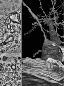
Researchers have mapped a tiny piece of the human brain in astonishing detail. The resulting cell atlas, which was described in Science and is available online, reveals new patterns of connections between brain neurons, as well as cells that wrap around themselves to form knots, and pairs of neurons that are almost mirror images of each other.

Posted in Uncategorized | Comments Off on Cubic millimetre of brain mapped in spectacular detail

Multiple sclerosis (MS) is a global health concern affecting around 2.6 million people. It is characterised by neural inflammation, myelin breakdown and cognitive decline. Cognitive impairment, especially reduced cognitive processing speed (CPS), which affects up to 67% of MS patients and frequently manifests before mobility concerns, is one of the disease’s most serious side effects. Effective adaptation and the application of cognitive rehabilitation treatments depend on the early diagnosis of cognitive impairment. Although pharmaceutical therapies have some drawbacks, endurance training has become a promising alternative. Intensity-controlled endurance exercise has the ability to delay the onset of MS symptoms and enhance cognitive function. Exercise has also been shown to have neuroprotective effects in a number of neurological disorders, including MS, Parkinson’s disease and stroke. This includes both aerobic and resistance training. A mix of aerobic exercise and weight training has shown promise, especially for people with mild cognitive impairment, but according to recent studies any amount of physical activity is beneficial to cognitive performance. In conclusion, this in-depth analysis highlights the crucial part endurance exercise plays in treating MS-related cognitive impairment. It improves not only neurological health in general but also cognitive performance. Exercise can help control MS in a way that dramatically improves quality of life and well-being.

Zameer U, Tariq A, Asif F, Kamran A. Empowering Minds and Bodies: The Impact of Exercise on Multiple Sclerosis and Cognitive Health. Ann Neurosci. 2024 Apr;31(2):121-123. doi: 10.1177/09727531241227674. Epub 2024 Feb 12. PMID: 38694717; PMCID: PMC11060125.

https://pubmed.ncbi.nlm.nih.gov/38694717/

Posted in Uncategorized | Comments Off on The Impact of Exercise on Multiple Sclerosis and Cognitive Health

SCH23390 is a widely used D1 dopamine receptor (D1R) antagonist that also elicits some D1R-independent effects. We previously found that the benzazepine, SKF83959, an analog of SCH23390, produces positive allosteric modulation of the Sigma-1 receptor (Sig1R). SCH23390 does not bind to the orthodoxic site of Sig1R but enhances the binding of 3H (+)-pentazocine to Sig1R. In this study, we investigated whether SCH23390 functions as an allosteric modulator of Sig1R. We detected increased Sig1R dissociation from binding immunoglobulin protein (BiP) and translocation of Sig1R to the plasma membrane in response to SCH23390 in transfected HEK293T and SH-SY5Y cells, respectively. Activation of Sig1R by SCH23390 was further confirmed by inhibition of GSK3β activity in a time- and dose-dependent manner; this effect was blocked by pretreatment with the Sig1R antagonist, BD1047, and by knockdown of Sig1R. SCH23390 also inhibited GSK3β in wild-type mice but not in Sig1R knockout mice. Finally, we showed that SCH23390 allosterically modulated the effect of the Sig1R agonist SKF10047 on inhibition of GSK3β. This positive allosteric effect of SCH23390 was further confirmed via promotion of neuronal protection afforded by SKF10047 in primary cortical neurons challenged with MPP+. These results provide the first evidence that SCH23390 elicits functional allosteric modulation of Sig1R. Our findings not only reveal novel pharmacological effects of SCH23390 but also indicate a potential mechanism for SCH23390-mediated D1R-independent effects. Therefore, attention should be paid to these Sig1R-mediated effects when explaining pharmacological responses to SCH23390.

Zhang GF, Zhu KL, Li Q, Zhang Y, Waddington JL, Du XD, Zhen XC. The classical D1 dopamine receptor antagonist SCH23390 is a functional sigma-1 receptor allosteric modulator. Acta Pharmacol Sin. 2024 Apr 11. doi: 10.1038/s41401-024-01256-1. Epub ahead of print. PMID: 38605179.

https://pubmed.ncbi.nlm.nih.gov/38605179/

Posted in Uncategorized | Comments Off on The classical D1 dopamine receptor antagonist SCH23390 is a functional sigma-1 receptor allosteric modulator

Abstract: Although B cells are implicated in multiple sclerosis (MS) pathophysiology, a predictive or diagnostic autoantibody remains elusive. In this study, the Department of Defense Serum Repository (DoDSR), a cohort of over 10 million individuals, was used to generate whole-proteome autoantibody profiles of hundreds of patients with MS (PwMS) years before and subsequently after MS onset. This analysis defines a unique cluster in approximately 10% of PwMS who share an autoantibody signature against a common motif that has similarity with many human pathogens. These patients exhibit antibody reactivity years before developing MS symptoms and have higher levels of serum neurofilament light (sNfL) compared to other PwMS. Furthermore, this profile is preserved over time, providing molecular evidence for an immunologically active preclinical period years before clinical onset. This autoantibody reactivity was validated in samples from a separate incident MS cohort in both cerebrospinal fluid and serum, where it is highly specific for patients eventually diagnosed with MS. This signature is a starting point for further immunological characterization of this MS patient subset and may be clinically useful as an antigen-specific biomarker for high-risk patients with clinically or radiologically isolated neuroinflammatory syndromes.

Zamecnik CR, Sowa GM, Abdelhak A, Dandekar R, Bair RD, Wade KJ, Bartley CM, Kizer K, Augusto DG, Tubati A, Gomez R, Fouassier C, Gerungan C, Caspar CM, Alexander J, Wapniarski AE, Loudermilk RP, Eggers EL, Zorn KC, Ananth K, Jabassini N, Mann SA, Ragan NR, Santaniello A, Henry RG, Baranzini SE, Zamvil SS, Sabatino JJ Jr, Bove RM, Guo CY, Gelfand JM, Cuneo R, von Büdingen HC, Oksenberg JR, Cree BAC, Hollenbach JA, Green AJ, Hauser SL, Wallin MT, DeRisi JL, Wilson MR. An autoantibody signature predictive for multiple sclerosis. Nat Med. 2024 Apr 19. doi: 10.1038/s41591-024-02938-3. Epub ahead of print. PMID: 38641750.

https://pubmed.ncbi.nlm.nih.gov/38641750/

Posted in Uncategorized | Comments Off on An autoantibody signature predictive for multiple sclerosis